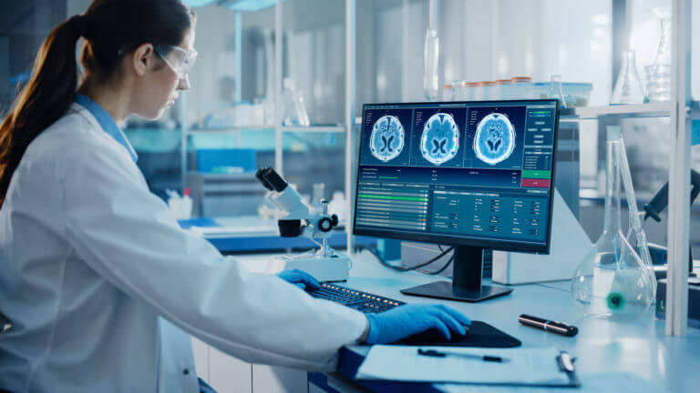
Neuroimaging is the process of producing images of the brain with the use of imaging techniques. Advancements in neuroimaging allow doctors to study the brain and its function and play a crucial role in identifying abnormalities associated with neurological conditions.
Brain injuries, spinal cord tumors, strokes, and aneurysms are all evaluated with the help of neuroimaging techniques. In this article, we talk about two of the most commonly used neuroimaging techniques employed by neurologists to manage their patients’ conditions.
At the end of the article, we also recommend a reputable neurology clinic in Southeast Michigan renowned for their advanced treatment of neurologic conditions.
MRI (Magnetic Resonance Imaging)
MRI is a noninvasive imaging technique that uses a powerful magnet and radio waves to generate highly detailed images of the brain. An MRI scan lets doctors visualize the different regions of the brain and detect abnormalities.
An MRI can be used to diagnose:
- Alzheimer’s disease and dementia
- Multiple sclerosis
- Spinal cord disorders
- Aneurysms
- Tumors
Here are some key aspects of MRI:
-
Structural Imaging
MRI excels at capturing intricate images of the brain’s anatomy. It can reveal structural changes in the brain, such as lesions, which is typical in neurologic conditions such as Alzheimer’s disease.
-
Brain Volumes
By measuring brain volumes, which is a measure of size or volume of the different brain structures. Magnetic resonance imaging allows doctors to measure gray matter, white matter, and regional brain volumes such as the hippocampus and amygdala. Changes in brain volumes are useful in assessing the impact on aging and neurodegeneration of the brain.
-
White Matter Integrity
Utilizing diffusion-weighted techniques, MRI can evaluate the integrity and connectivity of white matter tracts. White matter is what facilitates communication in the different regions of the brain. Reduced connectivity due to demyelination or axonal damage can be revealed in an MRI.
Some advancements in MRI used in brain imaging include:
-
Higher Image Resolution
Researchers have developed techniques to enhance the resolution of MRI scans. This allows for sharper and more detailed images of the brain, enabling doctors to visualize microscopic structures and abnormalities with greater precision.
-
Faster Scanning
Advancements in MRI software and hardware have led to faster scanning times. This reduces patient discomfort and improves efficiency in clinical settings.
-
Functional MRI (fMRI)
Functional MRI is a technique that measures changes in blood flow in the brain to map brain activity. It provides insights into how different regions of the brain function and interact with each other, aiding in the study of cognitive processes and neurological disorders.
-
Diffusion Tensor Imaging (DTI)
DTI is a specialized MRI technique that measures the movement of water molecules in brain tissue. It helps visualize the structural connections between different regions of the brain, allowing researchers to study neural pathways and identify abnormalities in conditions like traumatic brain injury and neurodegenerative diseases.
-
High-Field MRI
Increasing the strength of the magnetic field used in MRI scanners, such as 7 Tesla (7T) systems, improves image quality and enables better visualization of fine anatomical details. However, the use of high-field MRI requires careful consideration due to safety concerns and cost implications.
EEG (Electroencephalography)
Electroencephalography records the brain’s electrical activity using electrodes placed on the scalp. It measures voltage fluctuations resulting from neural activity and provides insights into brain function and activity.
Electroencephalography is used in the evaluation of certain neurological conditions, such as:
- Epilepsy
- Encephalitis or inflammation of the brain
- Brain injuries
- Stroke
Here are some things EEG can help study:
-
Brain Rhythms and States
Electroencephalography is particularly valuable in studying brain rhythms and states, from alpha to gamma waves. These patterns change based on cognitive tasks and sleep stages, as well as neurological conditions. Neurology specialists use EEG to diagnose and monitor epilepsy and sleep disorders, which are characterized by abnormal brain rhythms.
-
Event-Related Potentials (ERPs)
EEG can detect transient electrical responses in the brain known as ERPs. These responses are caused by specific stimuli or cognitive tasks. With EEG, the neurology doctor gains important information about a patient’s cognitive processing, memory, and sensory perception.
-
Epilepsy Localization
In individuals with epilepsy, electroencephalography plays a vital role in localizing the source of epileptic activity. By analyzing abnormal electrical discharges’ patterns and locations, EEG helps identify the origin of seizures. All in all, it is very helpful in guiding a patient’s treatment.
It’s truly remarkable how magnetic resonance imaging and electroencephalography contribute invaluable insights into the complex workings of the brain. It not only aids in the diagnosis and treatment of issues affecting the brain but is also used in ongoing research of these conditions.
MRI Imaging and EEG in Farmington Hills, Novi, and Howell, MI
If you are looking for high quality neurological treatments in Southeast Michigan, choose Associates in Neurology. Our team specializes in comprehensive, patient-centric treatments for neurological disorders. We make the latest advancements in neuroimaging available for our patients and continue to innovate for better patient outcomes.
To schedule an appointment with one of our board-certified neurologists, call our office near you at (248) 478-5512 or use our online request form. We look forward to providing you with the highest quality neurological care in Southeast Michigan.


